13-05-2021
A Stereotaxic Atlas Of The Rat Brain Pdf


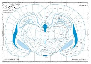
The hamster brain, we have chosen to follow closely the nomenclature used in the rat stereotaxic atlas of Paxinos and Watson [1998], the most widely-used atlas in rat neuroscience research. In this manner, we hope to facilitate communication between neuroscientists using rats and hamsters.

Stereotaxic Atlas Of The Brain
BOOK REVIEW
The Rat Brain in Stereotaxic Coordinates Authors: George Paxinos & Charles Watson Academic Press Australia, 1982 Softback, 1% pages (including 71 plates), 35.00 Aust $.
This is an excellent, large, well-labeled cytoarchitectonic atlas. Indeed it is the only complete rat brain atlas, depicting the brain from the olfactory bulb to the spinal cord. It is comprised of a brief introduction followed by 81 high quality photomicrographs of serial coronal, sagittal, and horizontal brain sections plus line drawings of these same sections. The sections are at 0.5 mm intervals and since they are in three planes they enable one to picture structures and localize them accurately in three dimensions. The stereotaxic coordinates have been carefully established and the distances from two reference points (the bregma and interauricular line) are indicated on each coronal plate. The major nuclei and fiber tracts are clearly illustrated and labeled. It is particularly helpful that the abbreviations used on any given plate are defined in full at the bottom of the page on which that plate appears. Separate lists of structures and abbreviations are also provided at the beginning of the volume. The atlas is for brains of 250-350 g male rats. This is valuable since the other well-established stereotaxic map is for the adolescent rat (150 g body weight). Nevertheless, a map for the 200-300 g rat would have been better in my opinion. The nomenclature and abbreviations used in the atlas deserve discussion. Unfortunately, no generally accepted neuroanatomical nomenclature exists, and the names of structures in common use are ambiguous. Nevertheless, the authors' choice of nomenclature is not aways the best. A number of terms and abbreviations in the atlas are rather unconventional. Despite this minor criticism, I highly recommend this atlas to neuroscientists, especially those who perform rat brain surgery. It will also be useful for non-neuroanatomists who need a good topographical guide to the rat brain. It is a worthwhile supplement to the rat stereotaxic atlases already available, and is among the best, if not the best, in its class.
Miklos Palkovits
NEUR-
E
319